
introduce
Eccrine duct fibroadenoma (ESFA) is a rare adnexal tumor characterized by eccrine ductal differentiation and a wide range of clinical manifestations.1 Histologically, ESFA is characterized by the presence of interconnected epithelial cell strands embedded within a fibrovascular stroma.2 Although ESFA has unique histological features, its clinical manifestations are nonspecific and variable. In this case report, we describe the clinical presentation of a female patient who presented with persistent erythema, scaling, and pin-sized blisters on her hands and feet, resembling the classic clinical features of dyshidrotic eczema. These symptoms persisted for several years, and reactive ESFA was diagnosed based on comprehensive clinical and histopathological examination.
case report
A 48-year-old Chinese woman presented with a 10-year history of erythema, scaling, and slowly expanding erythema on her hands and feet (Fig. 1a-d). She reported subjective itching and occasional pain, noting that the pain worsened with prolonged walking. Topical treatment with corticosteroid ointment and salicylic acid ointment failed to improve her symptoms. The patient denied any history of local trauma before the onset of the disease, and his family members had no similar experiences. Additionally, she had previously tested positive for hepatitis B antigen and had not received any treatment related to hepatitis B virus.
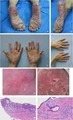 |
figure 1 Clinical, dermoscopic, and histological images taken at the patient’s initial visit. (arrive–d) The patient developed large, symmetrical erythema with desquamation of the palms, soles, and lateral edges of the skin. The patient’s feet were dry and cracked, and Auspitz’s sign was negative. Some fingernails and toenails showed deformities and obvious signs of damage. No similar skin lesions were found in other parts of the body. (and) Dermoscopy of the patient’s hands revealed a red background of the skin on the hands, with red areas and surrounding white scales. (F) Dermoscopy of the patient’s feet shows a light red heterogeneous background. Erosion appears as a creamy red area characterized by a central black scab-like area surrounded by concentric white circular lamellar scales. (G) Narrow epithelial cords connect to the skin, forming a grid-like structure (H and E × 50). (H) There is obvious typical duct differentiation locally (H and E×200). |
Fungal smear and culture tests of the toenails and skin lesions were negative. Dermoscopy of plantar lesions revealed clusters or nonspecific punctate blood vessels on a dark red background with yellowish-white scales (Fig. 1e–f). Subsequently, a biopsy was taken from the ingrown side of the left plantar, and histopathological examination revealed hyperkeratosis and interconnected chains of fine cuboidal cells extending from the epidermis into the upper dermis. Within these interconnected cell chains, small ductal structures and cystic changes were observed. No mitotic or developmental abnormalities were observed (Fig. 1g and h). Based on these findings, the diagnosis of reactive ESFA was established.
The patient was treated with topical mometasone furoate cream and mupirocin ointment, combined with zinc oxide and 20% cod liver oil ointment. Treatment involves applying a plastic wrap and bandage for 30 minutes, once a day. After 3 weeks, a significant improvement in the patient’s skin lesions was observed, characterized by reduced desquamation and erythema compared with the initial presentation (Fig. 2). The patient is currently being followed up, and no recurrence of skin lesions has been observed.
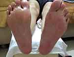 |
figure 2 The patient’s clinical manifestations after treatment. |
discuss
ESFA is a rare benign cutaneous adnexal tumor with eccrine apical duct differentiation.3 The age of the patients ranges from 16 to 80 years old, with the majority being 61 to 80 years old.4 The lesions mainly occur on the extremities, but can also be found on the back, abdomen, buttocks and other parts. Skin manifestations are diverse, including papules, plaques, nodules, etc., which can be isolated or multiple, symmetrical or linearly distributed.2 According to the Starink classification modified by French, there are five subtypes of eccrine fibroadenomas: solitary ESFA, multiple ESFA associated with edematous ectodermal dysplasia (Clouston and Schopf syndrome), and multiple ESFA without associated skin manifestations. ESFA (eccrine fibroadenomatosis), nonfamilial unilateral linear ESFA (nevi ES), and reactive ESFA associated with inflammatory or neoplastic processes.5.6 ESFA can occur alone or accompanied by skin ulcers, chronic lymphedema, amyloidosis, toenail trauma, burns, contusions, bullous pemphigoid, epidermolysis bullosa and other lesions. It may even be associated with or secondary to squamous cell carcinoma.7 The nature of ESFA, whether reactive or neoplastic, remains controversial, and it is considered a heterogeneous group of lesions. In this case, the patient denied dental aplasia, hypotrichosis, nail dystrophy, palmoplantar keratosis, and periorbital and lid margin apocrine cysts. In addition, there was no history of similar cases in the patient’s family. Based on the duration, clinical features, and histological appearance of the lesions, the patient was diagnosed with reactive eccrine duct fistula associated with dyshidrotic eczema. The occurrence of reactive ESFA may be related to long-term friction of hands and feet, sweat stimulation, and repeated sweat blisters.
Treatment of ESFA depends on the number, location, and resectability of the lesions. Surgical resection is preferred for a single small skin lesion, while multiple ESFA require individualized treatment options, such as cryotherapy, CO2 Laser, curettage, electrocautery, photodynamic therapy, localized radiation therapy, topical 5-fluorouracil, imiquimod ointment, and topical or systemic retinoids.8.9 However, reactive ESFA generally do not undergo malignant transformation. Some scholars believe that symptomatic treatment and regular follow-up are sufficient to control reactive ESFA. Therefore, treatment of reactive ESFA may not require surgical resection.10.11 In this case, the patient presented with eccrine duct fibroadenoma and dyshidrosis, indicating the presence of reactive ESFA. The lesions are extensive and difficult to remove. The patient was treated with topical corticosteroid ointment, mupirocin ointment, and cod liver oil and zinc oxide oil to treat dry and fissured areas of palmar and plantar erythema. The patient’s clinical symptoms improved and the skin lesions did not progress. Our case supports the notion that symptomatic treatment of the primary lesion can effectively control diffuse reactive ESFA. However, because of the diverse presentation of reactive ESFA lesions, clinicians often face challenges in diagnosing the lesions at the initial visit. Therefore, a comprehensive pathological examination (such as dermoscopy, fungal smear and culture, and relevant serology) is required.
Data Sharing Statement
All data used in this work are publicly available.
moral approval
The hospital ethics committee agreed to report this case.
Statement of consent
Formal written consent was obtained from the patient for publication of case details and associated images.
Acknowledgments
We thank Dr. Xin Zhou for valuable suggestions and discussions.
Author contributions
All authors made a substantial contribution to the reported work, whether in conception, study design, execution, acquisition of data, analysis, and interpretation, or in all these areas; participated in drafting, revising, or critically reviewing the article; final approval the version to be published; have agreed on the journal to which the article will be submitted; and agree to be accountable for all aspects of the work.
funds
This research was funded by the Guangzhou Science and Technology Planning Project (Grant No.: 2023A03J0468).
Disclosure
The authors declare that there are no conflicts of interest in this work.
refer to
1. Takeda H, Mitsuhashi Y, Hayashi M, Kondo S. Eccrine syringofibroadenoma: case report and literature review. european journal of dermatology. 2001;15(2):147–149. doi:10.1046/j.1468-3083.2001.00213.x
2. Temnithikul B, Jerasutus S, Sudtikoonaseth P, Voravutinon N, Kootiratrakarn T, Kattipathananpong P. Eccrine syringofibroadenoma (ESFA): a report of two cases. Dermatol Practice Concept. 2016;6(1):5–8. doi:10.5826/dpc.0601a03
3. Tiwari AK, Firdous J, Mishra DK, Chaudhry SS. A case report of reactive solitary eccrine syringofibroadenoma. Indian Dermatol Online J. 2017;8(1):35–38. Number: 10.4103/2229-5178.198766
4. Botino CB, Guimarães TF, Gomez FR, Am D, Lima RB, Martins CJ. Solitary eccrine duct fibroadenoma—a case report. Bra Dermatol. 2015;90(3Suppl1):235–238. Number: 10.1590/abd1806-4841.20153802
5. Khan HA, Kumarasinghe P, Wood B. Delayed eccrine duct fibroadenoma of the feet in a patient with hypohidrotic ectodermal dysplasia. australian journal of dermatology. 2021;62(3):383–385. doi:10.1111/ajd.13604
6. Chukwuma O, Walker A, Motaparthi K, Montanez-Wiscovich M. Refractory erosions on the palms and soles: a rare manifestation of eccrine fibroadenoma. JAAD Case Representative. 2020;6(7):590–592. doi:10.1016/j.jdcr.2020.05.004
7. Lee JS, Park H, Yoon HS, Cho S. Eccrine fibroadenoma associated with Bowen disease: case report and literature review. Ann DeMarto. 2020;32(1):57–63. doi:10.5021/ad.2020.32.1.57
8. Sirikham T, Rojhirunsakool S, Vachiramon V. Reactive eccrine fibroadenoma associated with neuropathy, venous stasis, and diabetic foot ulcers. Case Representative Dermatol. 2016;8(2):124–129. Number: 10.1159/000446469
9. Morganti AG, Martone FR, Macchia G, et al. An unusual manifestation of radiotherapy for eccrine fibroadenoma. dermatotherapy. 2010;23(Suppl 1):S20–3. doi:10.1111/j.1529-8019.2009.01282.x
10. Zhou Ping, Li Feng, Liu Hong, Wang L. Solitary eccrine fibroadenoma on the dorsum of the hand. indian journal of dermatology. 2020;65(6):560–561. doi:10.4103/ijd.IJD_331_18
11. Sugita Y, Makino T, Matsui K, Shimizu T. Reactive eccrine syringofibroadenoma on the heel, clinically similar to squamous cell carcinoma. Case RepresentativeDermatol Med. 2019;2019:4735739. Number: 10.1155/2019/4735739