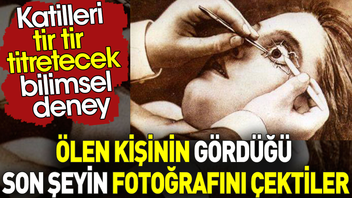
With the idea introduced in the seventeenth century, an attempt was made to photograph the last things seen by the dead.
According to a popular article on Sour Things, in the seventeenth century, scientists tried to extract an image that formed on the retinas of the eyes of dead people. This was also called optography.
Basically, the word Optography comes from a combination of two Greek words. Optics: means vision or eye, and chart means something related to the workspace or writing.
We can say that scientists first put forward this idea in the seventeenth century. optics; They defined the optogram, which is the image formed on the retina, as the process of viewing or receiving.
According to this idea, scientists at the time believed that it was possible for the eye to capture our last vision at the moment of death. Of course, we can say that this idea emerged as a more dominant idea after the invention of cameras and photography.
In 1876, a German physiologist named Franz Christian Paul discovered rhodopsin, a light-sensitive protein found in the rod cells of the retina that functions just like the nitrates in a camera plate. When this detected protein was exposed to light, bleaching occurred.
Because Paul’s life ended early, he was unable to see the impact his work had. Later, another German physiologist, Wilhelm Kuehne, followed up on this theory and began conducting various experiments.
In his experiments, he used a large number of animals. He removed the animals’ eyes immediately after death and exposed them to various chemical substances to fix the image on the retina. After these experiments, he discovered that the best substance was potassium alum, which is a type of alum. We call salt alum.
A rabbit was tied with its head facing a barred window. From this position, the rabbit could only see the window and the cloudy sky. The animal’s eyes were covered with a cloth for several minutes to acclimate it to the dark, i.e. to allow rhodopsin to accumulate in its rods. The animal was then exposed to light for three minutes.
He was then immediately beheaded and his eyes removed. An incision is made in the removed eyes, and the back half of the eyeball containing the retina is removed and immersed in an alum solution for fixation.
The next day, Kuehne saw an image of a window with a clear pattern of bars imprinted on the retina with bleached, unmodified rhodopsin:
After these studies, Kuhne wanted to conduct his experiments on humans, so in 1880, a death row prisoner named Erhard Gustav Reif had his eyes removed after his execution and was delivered to Kuhne’s laboratory at the University of Heidelberg.
Years later, in 1975, the Heidelberg police wanted to re-examine Kuhne’s experiments and results using modern scientific techniques, updated information and advanced equipment. For this reason, they turned to the expertise of Evangelos Alexandridis from Heidelberg University. In a manner reminiscent of Kuehne, Alexandridis was able to create many different high-contrast images from rabbits’ eyes. But he concluded that optics had no potential as a forensic tool.
(Tags for translation)killer

