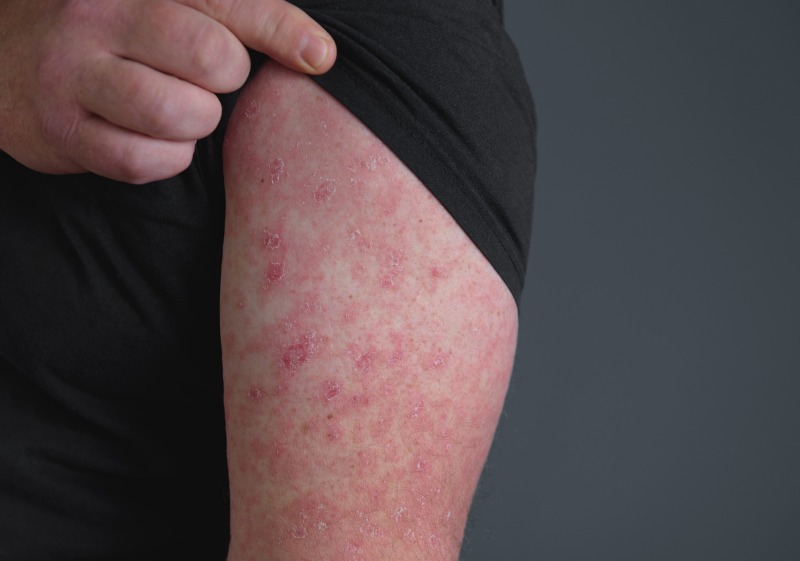
Distinctive features of 4 severe drug eruptions.
Adverse drug reactions are the fifth leading cause of death among all diseases, accounting for 5% to 10% of hospitalizations worldwide. They remain a challenge in modern healthcare, especially as complications and treatments become increasingly complex.
By definition, adverse drug reactions are unexpected harmful events that occur as a result of the use of drugs in clinical practice. They are associated with prolonged hospital stays, increased readmissions and costs of care, and death. 30% to 45% involve the skin. Risk factors include female gender, older age, more frequent medication use, immunocompromise, and autoimmune diseases.
Identifying the type of rash caused by a drug can be difficult. Clinicians are familiar with the clinical features of the two most common drug-induced skin reactions: morbilliform rash and urticaria:
• Morbilliform drug eruption, also called eruption or maculopapular eruption, is the most common and usually manifests as an erythematous maculopapular rash 1 to 2 weeks after drug exposure.
• Urticaria is the second most common rash and manifests as migrating, pruritic, annular patches that usually appear within hours of initial drug exposure.
Penicillins, sulfonamides, anticonvulsants, and nonsteroidal anti-inflammatory drugs (NSAIDs) are associated with higher rates of skin reactions. The latter and salicylates are more associated with urticaria than with morbilliform drug-induced rash. Severe drug rash is less common but can be life-threatening. They enable early identification, allowing suspected drugs to be immediately suspended.
| Stevens-Johnson syndrome/toxic epidermal necrolysis (SJS/TEN) |
SJS and TEN are overlapping disorders characterized by mucocutaneous reactions with epidermal necrosis and sloughing of the affected skin. Conditions are divided into 3 categories based on their severity and percentage of body surface area involved:
• SSJ: Lesion area <10%
• SSJ/NET overlay: Lesion area 10%~30%
• net: Lesion area >30%.
The global incidence of SJS/TEN is estimated to be 6 cases per million person-years in Europe and the United States. Rates are higher in adults, women, and Asians or blacks.
The most common triggering drugs are allopurinol, antibiotics (especially sulfonamides), antiepileptic drugs, and nonsteroidal anti-inflammatory drugs.
Immune checkpoint inhibitors are increasingly used to treat malignancies and are associated with severe drug eruptions, including SJS/TEN.
> Symptoms appear 1-3 weeks later
The rash usually appears 1 to 3 weeks after taking the medication. Lesions usually appear first on the face and chest and then spread symmetrically. They begin as macules with erythema and a dark necrotic center in the target lesion, which later turn into blisters, erosions, or ulcers with associated exfoliation. They usually have a positive Nikolsky sign, in which traction pressure causes shearing and erosion of the epidermis.
> Systemic manifestations
Systemic manifestations are common and include influenza-like symptoms, fever, lymphadenopathy, and mucosal involvement (conjunctiva, oropharynx, esophagus, and genitals). Up to 90% of patients experience this symptom. Mouth ulcers, grit in the eyes, painful swallowing, and difficulty urinating are common.
The most clinically important factor in mucosal involvement is the sequelae of mucosal ulceration, leading to scarring and stenosis, affecting multiple organ systems—the cornea, urethra, esophagus, and pulmonary tract.
this serious complications SJS/TEN includes respiratory failure, shock, functional capacity deficit, and infection. The average mortality rate for SJS is 1% to 5% and for TEN is 25% to 35%.
> diagnosis
The diagnosis of SJS/TEN is based on a history of drug exposure and clinical evidence of typical mucocutaneous lesions. The gold standard for diagnosis is skin biopsy combined with conventional histopathology and direct immunofluorescence studies.
If the diagnosis is uncertain, biopsy may be useful even in the early stages, but will be more definitive in later stages, when the distinctive appearance of full-thickness necrosis and subepidermal detachment is already present.At this stage, subsequent biopsy can help rule out SJS/TEN-like diagnoses such as staphylococcal squamous skin syndrome and other systemic blistering rashes such as exfoliative erythroderma, bullous pemphigoid, pemphigus sore ordinary and linear immunoglobulin A dermatoses.
> Supportive measures and timely referrals are important.
The first and most important step in the management of patients with SJS/TEN is immediate identification and discontinuation of the suspect medication.
Immediate discontinuation of the pathogen before erosions and blisters develop means a reduced risk of death. The SCORTEN (Severity of Disease in Toxic Epidermal Necrolysis) tool, which includes prognostic indicators such as heart rate, age and kidney function, can be used to determine a patient’s risk of death with SJS/TEN.
The mainstay of treatment is supportive: intravenous fluids, electrolyte replacement, nutritional support, pain control, and infection prevention. Regular skin and blood cultures can help detect and treat superinfections. Immediate referral to burn units and specialists (e.g. ophthalmology, urology) depending on organ involvement.
The treatment method is corticosteroids This is controversial, but intravenous immune globulin alone or in combination with corticosteroids has shown varying degrees of success. Other options include plasmapheresis, immunosuppressants (cyclosporine, cyclophosphamide, thalidomide) or various combinations of these options, as well as any of the above treatments. Prophylactic systemic antibiotics should be avoided unless infection studies raise concerns about bacterial superinfection.
| Drug reaction with eosinophilia and systemic symptoms (DRESS) |
DRESS (Drug Reaction with Eosinophilia and Systemic Symptoms) is a delayed multiorgan reaction that usually occurs 2 to 6 weeks after starting a medication, although certain drugs, such as antibiotics, may cause the rash to appear earlier.
Its incidence ranges from 1 in 1,000 to 1 in 10,000 drug exposures, and it is responsible for nearly 18% of adverse drug reactions affecting the skin in hospitalized patients. The most common adverse drugs include antiepileptic drugs (carbamazepine, phenytoin, lamotrigine, phenobarbital), allopurinol, sulfonamides (sulfasalazine, dapsone, trimethoprim- sulfamethoxazole), minocycline, vancomycin, and antituberculosis drugs (isoniazid, rifampicin, ethambutol, pyrazinamide).
| severity score Toxic epidermal necrolysis (SCORTEN) | |
| SCOTEN parameters | Fraction |
| Age ≥ 40 years old | 1 |
| malignant tumor | 1 |
| Heart rate >120/min | 1 |
| Initial dermal detachment rate >10% | 1 |
| Uremia>65 mg/dl | 1 |
| Blood glucose >252 mg/dL | 1 |
| Bicarbonate ≥20 mEq/l | 1 |
| Total Score | Predicted percent risk of death |
| 0-1 2 3 4 >5 |
3.2 12.1 35.8 58.3 90 |
> Fever, rash, facial edema, eosinophilia
DRESS typically begins with fever and a severe, itchy, nonspecific rash affecting more than 50% of the body’s surface area. Patients typically present with central facial edema but sparing of the periorbita. The rash is usually maculopapular, but the lesions are polymorphic and may appear as plaques, vesicles, target lesions, urticaria, exfoliation, eczema, or rarely, lichenoid rashes.
In addition to rash and fever, other manifestations may include lymphadenopathy, hematological abnormalities, and internal organ involvement (usually the liver, kidneys, lungs, and heart). Up to 95% of patients with DRESS have eosinophilia. During the long-term clinical course, sequential reactivation of several human herpesviruses (particularly types 6 and 7), and less commonly infections with Epstein-Barr virus and cytomegalovirus, can be observed.
The course of the disease may wax and wane with multiple outbreaks. The average mortality rate is 4% to 10% due to multiorgan failure (most commonly hepatic necrosis), and long-term complications include exfoliative dermatitis, acute necrotizing eosinophilic myocarditis, and autoimmune sequelae such as thyroid disease, vitiligo, and alopecia. Aretalupus erythematosus, autoimmune hemolytic anemia, and fulminant type 1 diabetes.
> RegiSCAR: Diagnostic Criteria Resource
Clinical findings of rash involving visceral organs and eosinophilia should raise suspicion of DRESS and initiate appropriate studies. The RegiSCAR (Registrar of Serious Cutaneous Adverse Reactions) criteria are the most detailed and commonly used diagnosis. Follow-up blood tests should be performed based on the organ suspected to be affected.
The histopathology of DRESS is nonspecific and includes spongiosis, basal vacuolation, necrotic keratinocytes, dermoepidermal infiltrates, dermal edema, and perivascular lymphocytic infiltration with or without eosinophils.
Identifying the causative agent can be challenging due to the late onset of disease after drug exposure.method in vitro The most reliable method of confirming drug causation is the lymphocyte transformation test, which is particularly useful during anticonvulsant and antituberculous therapy. The test assesses activation of drug-specific T cells with a sensitivity of 73% and a specificity of 82%, but must be performed 2 to 6 months after the acute phase.Skin test in vivoparticularly in those treated with patches and later intradermal treatments, may also be helpful in identifying the causative agent.
> multidisciplinary management
Management of DRESS requires a multidisciplinary approach depending on the organs affected and severity. If the patient’s disease is mild and the aminotransferase levels are moderately elevated (<3 times the upper limit of normal), topical corticosteroids can be used for symptomatic treatment.
The preferred treatment for severe disease is systemic corticosteroids at moderate to high doses.
For patients who do not respond to corticosteroids, intravenous immunoglobulins and Janus kinase inhibitors can be used with some success. Other alternatives include immunosuppressants (cyclophosphamide, cyclosporine, interferon, mycophenolate mofetil, rituximab), antiviral drugs, and plasmapheresis. Antibiotics and antipyretics should be avoided unless there is clear evidence of infection.

